Significance Statement
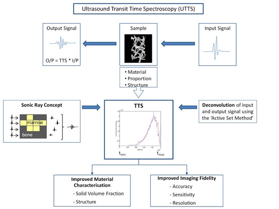
It has previously been proposed [1] that ultrasound propagation in complex porous composites such as cancellous bone may be described by a concept of parallel sonic rays, the transit time of each ray defined by the proportion of bone and marrow propagated, a minimum (tmin) solely through bone and a maximum (tmax) solely through marrow [2]. A Transit Time Spectrum (TTS) plots the proportion of sonic rays having a particular transit time, the TTS is therefore determined by the ultrasound velocities of the two constituent materials, their relative proportion, and the structure of the sample.
Considering ultrasound measurement of a test sample, we may mathematically describe the output signal as the convolution of the input signal and the sample’s TTS. Conversely, the TTS may be derived by deconvolution of the experimental output and input signals, as described in the highlighted paper.
We envisage that ultrasound transit time spectroscopy (UTTS) offers significant potential to improve both material characterisation of complex porous solids and the fidelity of ultrasound imaging.
From the material characterisation perspective, we have recently tested the hypothesis that UTTS may reliably estimate the solid volume fraction of simplified bone:marrow replica models; agreement between true (geometric calculation) with both predicted (computer simulation) and experimentally-derived values were both above 95%. Current work is testing the hypothesis that cancellous bone structure may be estimated from the profile of a transit time spectrum.
Noting that the fidelity of ultrasound imaging may be improved with increased accuracy and precision of transit time measurement, we have recently compared the ability of a deconvolution-derived TTS and the established technique of matched-filtering to identify signal locations that have been lost due to phase interference; deconvolution was shown to be as accurate as the established matched-filtering approach, also providing improved side-lobe suppression [3].
- Langton C M; 2011; 25th Anniversary of BUA for the Assessment of Osteoporosis – Time for a New Paradigm?; Engineering in Medicine; 225 (2),113-125
- Langton C M & Wille M-L; 2013; Experimental and computer simulation validation of ultrasound phase interference created by lateral inhomogeneity of transit time in replica bone:marrow composite models; Engineering in Medicine; 227(8), 890-895
- Wille M-L, Zapf M, Ruiter N V, Gemmeke H, and Langton C M; 2014; Comparison of Active-Set Method Deconvolution and Matched-Filtering for Derivation of an Ultrasound Transit Time Spectrum; Proc 5th Biomedical Engineering Conference, Vietnam
Proc Inst Mech Eng H. 2014 Apr;228(4):321-9.
Langton CM, Wille ML, Flegg MB.
Biomedical Engineering & Medical Physics Discipline, Science & Engineering Faculty and Institute of Health & Biomedical Innovation, Queensland University of Technology, Brisbane, QLD, Australia.
Abstract
The acceptance of broadband ultrasound attenuation for the assessment of osteoporosis suffers from a limited understanding of ultrasound wave propagation through cancellous bone. It has recently been proposed that the ultrasound wave propagation can be described by a concept of parallel sonic rays. This concept approximates the detected transmission signal to be the superposition of all sonic rays that travel directly from transmitting to receiving transducer. The transit time of each ray is defined by the proportion of bone and marrow propagated. An ultrasound transit time spectrum describes the proportion of sonic rays having a particular transit time, effectively describing lateral inhomogeneity of transit times over the surface of the receiving ultrasound transducer. The aim of this study was to provide a proof of concept that a transit time spectrum may be derived from digital deconvolution of input and output ultrasound signals. We have applied the active-set method deconvolution algorithm to determine the ultrasound transit time spectra in the three orthogonal directions of four cancellous bone replica samples and have compared experimental data with the prediction from the computer simulation. The agreement between experimental and predicted ultrasound transit time spectrum analyses derived from Bland-Altman analysis ranged from 92% to 99%, thereby supporting the concept of parallel sonic rays for ultrasound propagation in cancellous bone. In addition to further validation of the parallel sonic ray concept, this technique offers the opportunity to consider quantitative characterisation of the material and structural properties of cancellous bone, not previously available utilising ultrasound.