Significance Statement
Multiphoton microscopy has experienced numerous technological advancements and found many applications in biomedical fields including embryology, oncology and neuroscience. As opposed to 1-photon excitation, the implementation of near infrared excitation light in multiphoton microscopy effectively suppresses tissue scattering enabling deeper penetration. In view of imaging biological tissues, tissue scattering and absorption of water should be kept low, which depends on prudent selection of excitation wavelength.
Tissue scattering as well as water absorption results in exponential decay of excitation light as imaging depth increases. Recent experimental and theoretical studies have proven that the 1700-nm window is an optimum window for deep-tissue multiphoton microscopic such as brain [1]. However, the depth of imaging in multiphoton microscopy at the optimal window is limited by the available signal level deep within the tissue. A number of methods have been proposed for the enhancement of signal generation, for example, customized optics, excitation with a powerful laser, and optimal filling of the objective lens.
High numerical aperture immersion objective lenses are commonly used in multiphoton microscopy. At the 1700 nm window, the immersion medium could pose severe absorption problem even if the thickness is on the order of mm. For brain imaging, deuterium dioxide immersion has been proven to be more efficient in terms of signal generation compared with water immersion [2]. In contrast, the implementation of the multiphoton microscopy to diagnose skin diseases such as psoriasis and malignant melanoma, requires oil immersion to match the refractive index properly. Owing to lack of measured absorption spectrum at the optimal 1700-nm, which the immersion oil ought to be selected to allow for the deep penetration in skin is unknown.
Targeting deep skin imaging excited at the 1700-nm window, Shenzhen University researchers led by Professor Ke Wang and Professor Ping Qiu demonstrated accurate measured spectra of a number of commonly used immersion oil to determine the absorption data for the 1700-nm window. They also proposed the exact wavelength that was to be used within this optimal window for the highest signal level. Finally, they demonstrated second-harmonic generation as well as third harmonic generation imaging of a mouse tissue in order to corroborate their selection of oil and excitation wavelength at the 1700-nm window. Their work is published in Optics Express [3].
The authors used cuvettes with shorter and thicker immersion medium thickness as reference and sample respectively. They obtained the accurate absorption spectrum data by computing the ratio of the sample absorption data and the reference absorption data. This approach helped them eliminate measurement error arising from surface reflection or intrinsic absorption of the quartz cuvettes.
The research team obtained spectra of three commercially available immersion oils and glycerol from 1200 nm to 1900 nm, which covered both the 1700nm and 1300nm windows, for deep tissue multiphoton microscopy. Their results showed that that the absorption spectra of the three immersion oils were quite similar in magnitude and shape, despite the fact that they were of varying compositions.
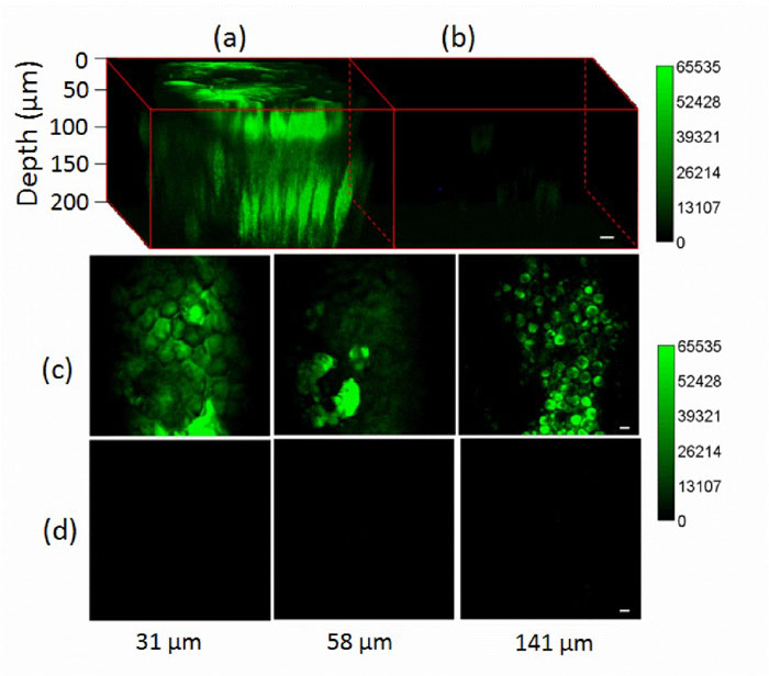
Based on the measured results, glycerol- heavy water mixture immersion indicated lower absorption than glycerol-water mixture immersion. For the oil immersion, within the optimal 1700nm window, 1600nm excitation was considered owing to the much smaller absorption by the immersion medium. Selecting ideal immersion oil and excitation wavelength was of ultimate importance to deep tissue multiphoton microscopy. The authors demonstrated the significance of immersion oil and excitation wavelength selection, by comparing skin imaging at both 1600 nm and 1700 nm. Their results clearly indicated that 1600-nm excitation lead to order-of-magnitude higher signal levels with even less excitation power before the immersion oil, compared with 1700-nm excitation. They expected this would help increase penetration in skin imaging in future experiments.


References
[1] N. G. Horton, K. Wang, D. Kobat, C. G. Clark, F. W. Wise, C. B. Schaffer, and C. Xu, “In vivo three-photon microscopy of subcortical structures within an intact mouse brain,” Nat. Photonics 7(3), 205-209 (2013).
[2] Y. X. Wang, W. H. Wen, K. Wang, P. Zhai, P. Qiu, and K. Wang, “Measurement of absorption spectrum of deuterium oxide (D2O) and its application to signal enhancement in multiphoton microscopy at the 1700-nm window,” Appl. Phys. Lett. 108(2), 021112(2016).
[3] Ke Wang, Wenhui Wen, Yuxin Wang, Kai Wang, Jiexing He, Jiaqi Wang, Peng Zhai, Yanfu Yang, And Ping Qiu. Order-of-magnitude multiphoton signal enhancement based on characterization of absorption spectra of immersion oils at the 1700-nm window. Vol. 25, No. 6 | 20 Mar 2017 | OPTICS EXPRESS 5909.
Go To Optics Express