Statement of Significance
Microfluidics and Nanofluidics, May 2014, Volume 16, Issue 5, pp 849-856.
Chihchen Chen, Bo-Ren Lin, Hsi-Kai Wang, Shu-Ting Fan, Min-Yen Hsu, Chao-Min Cheng.
Institute of Nanoengineering and Microsystems, National Tsing Hua University, Hsinchu, 300, Taiwan.
Taichung Veterans General Hospital, Taichung, 407, Taiwan.
Abstract
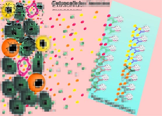
We present a paper-based immunoaffinity platform for separating subsets of extracellular vesicles (EVs). This method is inexpensive, robust, easy-to-use (easy-to-prepare), and compatible with downstream analyses, such as scanning electron microscopy (SEM), enzyme-linked immunosorbent assays (ELISA), and transcriptome analysis. Extracellular vesicles are small membranous vesicles shed from both healthy and diseased cells and contain nucleic acid and protein cargo. EVs have been increasingly recognized as a means of cell–cell communication and hold a great potential for clinical applications. However, current protocols for isolation of EVs are often lengthy, cumbersome and require expensive equipment. Here, we have isolated EVs from small volumes of both human serum and aqueous humor samples using paper-based immunoaffinity devices. Captured EVs were analyzed morphologically using SEM and appeared statistically different in size (p value < 2.4 × 10⁻²²) and circularity (p value < 3.6 × 10⁻⁹) between subsets of Extracellular vesicles bearing different surface markers. Assessing the amount of Extracellular vesicles captured using paper-based ELISA using antibodies conjugated to horseradish peroxidase to produce a colorimetric readout was accomplished within 10 min. RNAs contained in EVs can be extracted to provide information for disease management. These paper-based immunoaffinity devices, we believe, open opportunities for a wide range of applications in both basic biology and clinical medicine.