Significance Statement
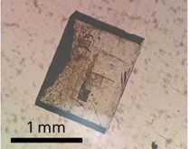
The indentation microfracture method is revisited here as a tool for evaluating surface toughness in bioceramics for artificial joint applications. Despite the experimental expediency of this method and its uniqueness in evaluating surface toughness, challenging tasks have historically been found to accompany the indentation procedure when the toughness evaluation is challenged at a fully quantitative level. Leaving aside empirical equations, which have been proposed by a number of different authors, we looked for a more rigorous way to extract surface toughness characteristics from the indentation method.
Our approach to quantitative assessments was built upon the well-established notion of (Vickers) crack opening displacement (COD) test. The improvement proposed here to this established procedure consisted in coupling it with quantitative assessments of microscopic stress fields by confocal (spatially resolved) Raman piezo-spectroscopy. Empowered by the Raman microprobe, the indentation micro-fracture method is shown capable of providing a reliable surface toughness evaluation in silicon nitride biomaterials. Different from bulk toughness, surface toughness is discussed here as a fundamental parameter in designing the microstructure of bioceramic bearing couples with improved tribological performance. The present spectroscopic development of the indentation COD technique paves the way to reliably compare the structural properties of bioceramic surfaces as distinct from their bulk properties, and to quantitatively monitor their modification upon environmental exposure.
We believe that the newly proposed Raman-assisted indentation toughness method could be both useful and innovative in the field of bioceramic bearings. Modern bioceramics for load-bearing applications in artificial hip joints possess refined microstructures, thoroughly optimized for the concurrent achievement of bioinertness, high structural reliability, and superior wear resistance. In commercial components, structural reliability is usually certified through statistically validated and standardized procedures, including assessments of fracture toughness, burst strength, and its related Weibull distribution, which thus deal with the bulk properties of the biomaterial.
On the other hand, the evaluation of local mechanical properties at the surface of biomaterials has so far less attention and yet lacks appropriate standardization. The main (only) standard evaluation consists of the analysis of wear resistance, performed through wear simulation testing under standardized conditions. However, advanced studies concerned with the way in vitro simulations are kinematically conducted during standard tests in comparison to in vivo conditions, have clearly shown the need to modify the existing standards. In particular, the importance was shown of including in the hip-joint simulation protocol a lateral shift in the contact during swing phase (usually referred to as “microseparation”). This additional (kinematical) feature serves to simulate an offset deficient head (or joint laxity), which represents the physical origin of the clinically relevant wear rates and enhanced patterns detected on retrievals, in contrast with the outputs of merely frictional wear simulations. In hard-on-hard bearings, the occurrence of microseparation between femoral head and acetabular cup, and the repeated micro-shock events that accompany it, have a profound impact on the micromechanical damage produced by the in vivo wear phenomenon.
From a micromechanical point of view, the occurrence of repeated impingements and micro-shock events (i.e., rather than the continuously lubricated sliding between the mating surfaces of the joint envisaged by standard hip simulation tests) has an impact on both the amount and type of surface damage that one should expect to occur during the lifetime of the joint components. In biomaterials development, designing bioceramic microstructures with high fracture toughness and/or introducing appreciable magnitudes of compression residual stresses into the joint surfaces are two key concepts in counteracting the occurrence of microseparation-related surface damage. In bearings made of oxide ceramics, larger amounts of damage as compared to surfaces merely subjected to frictional simulations has indeed been observed when microseparation was introduced in the gait simulation kinetics. Moreover, tougher bioceramics have shown, ceteris paribus, a lesser amount of surface damage.
Having clear in mind that brittle bioceramics are more prone to surface damage than the tough ones, one could also foresee a variety of tribochemical effects related to the biological environment, which might strongly affect the surface toughness of bioceramics and degrade it below the respective bulk values measured in the as-manufactured samples. Environmentally exposed components might thus progressively experience lower surface toughness values as compared to bulk values. Accordingly, toughness analyses at bioceramic surfaces necessarily require a specialized (local) evaluation method, different from those already standardized for bulk toughness.
In our work, we attempted to implement the indentation method to quantitatively assess surface toughness in silicon nitride bioceramics. We set the originality of our contribution here in the quantitative use of Raman piezo-spectroscopy in directly measuring and quantitatively deconvoluting the different stress fields associated with the indentation print. By doing so, an intrinsic (local) toughness value for crack initiation and its rising upon crack propagation could directly be extracted from experimental micro-stress data, rather than being indirectly deduced according to semi-empirical equations. Coupled with an appropriate sampling procedure to ensure statistical validity, the proposed method appears to improve, with respect to other indentation-related methods, the testing reliability in estimating surface toughness. Surface toughness could significantly differ from bulk toughness and represents a fundamental parameter in evaluating the potentiality of ceramic biomaterials with respect to their long-term tribochemical performance.

Journal Reference
J Mech Behav Biomed Mater. 2016 ;54:328-45.
Pezzotti G1, Enomoto Y2, Zhu W2, Boffelli M2, Marin E2, McEntire BJ3.
[expand title=”Show Affiliations”]- Ceramic Physics Laboratory, Kyoto Institute of Technology, Sakyo-ku, Matsugasaki, 606-8126 Kyoto, Japan. Electronic address: [email protected].
- Ceramic Physics Laboratory, Kyoto Institute of Technology, Sakyo-ku, Matsugasaki, 606-8126 Kyoto, Japan.
- AMEDICA Corporation, 1885 West 2100 South Salt Lake City, UT 84119, USA. [/expand]
Abstract
Indentation micro-fracture is revisited as a tool for evaluating the surface toughness of silicon nitride (Si3N4) bioceramics for artificial joint applications. Despite being unique and practical from an experimental perspective, a quantitative assessment of surface fracture toughness using this method is challenging. An improved method has been developed, consisting of coupling indentation with confocal (spatially resolved) Raman piezo-spectroscopy. Empowered by the Raman microprobe, the indentation micro-fracture method was found to be capable of providing reliable surface toughness measurements in silicon nitride biomaterials. In designing the microstructures of bioceramic bearing couples for improved tribological performance, surface toughness must be considered as a fundamentally different and distinct parameter from bulk toughness. The coupling of indention crack opening displacements (COD) with local stress field assessments by spectroscopy paves the way to reliably compare the structural properties of bioceramics and to quantitatively monitor their evolution during environmental exposure.
Copyright © 2015 Elsevier Ltd. All rights reserved.
Go To J Mech Behav Biomed Mater.