Significance Statement
Optical microscopy can be used for minimally invasive examination of living cells. However, the resolution of conventional light microscopy is restricted to ~200 nm (half the wavelength of visible light). This Abbe resolution limit has recently been overcome by several fluorescence-based super-resolution nanoscopy methods, one of them being stimulated emission depletion (STED) nanoscopy. This is a raster-scanning approach that is based on a confocal microscope. It offers improved spatial resolution by overlaying the excitation beam with a doughnut-shaped depletion beam that confines the fluorescence emission to the center of the excitation spot. The depletion beam thus features an intensity profile which is zero at the center of the Gaussian excitation spot.
Low intensity of the STED beam in the periphery of the confocal region may lead to incomplete depletion that appears as a low resolution background. The problem can be solved by increasing the power of the depletion beam. This, however, leads to low-resolution background due to STED beam re-excitation as well as photodamage in the high-power zones.
In a recent paper published in Nature Photonics, Professor G. Ulrich Nienhaus and his co-workers at the Karlsruhe Institute of Technology in Germany introduced the stimulated emission double depletion (STEDD) approach in a bid to selectively eliminate the low frequency background.
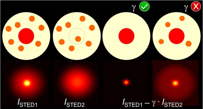
In the STEDD method, at each pixel location, the ‘normal’ STED beam is followed by a second, Gaussian-shaped STED2 beam with a specific time delay that eliminates the useful signal at the center so that only background excitation exists. Photons registered prior to and after the arrival of the STED2 beam are collected in two separate images. Thus, the image containing photons detected before the STED2 beam arrives contains the useful signal and the background. The image containing photons detected after the STED2 beam arrives includes only background. To obtain the high-resolution image, the authors calculate the weighted difference of photons assembled before and after the second pulse. The weight factor depends on the fluorescence lifetime of the fluorophores as well as the delay time between the first and the second pulse. The outcome of their technique is a highest resolution, background-free image.
This approach requires an approach that can reliably and precisely extract the weight factor used in the analysis of stimulated emission double depletion images from acquired data directly that has been reported by Professor G. Ulrich Nienhaus and his postdoctoral fellow Dr. Peng Gao in a second paper in Optics Letters. Their determination of the weight factor, which is based on maximization of the high spatial frequency components in the image, is a powerful method for differential background suppression approaches as employed in STED nanoscopy as well as focal modulation-based imaging.
References
Gao, P., Prunsche, B., Zhou, L., Nienhaus, K., & Nienhaus, G. Ulrich. Background Suppression in Fluorescence Nanoscopy with Stimulated Emission Double Depletion. Nature Photon. 11 (2017) 163-169.
Go To Nature Photonics
Peng Gao1,2 and G. Ulrich Nienhaus1,2,3,4. Precise Background Subtraction in Stimulated Emission Double Depletion Nanoscopy. Optics Letters Vol. 42, No. 4 / February 15 2017
[expand title=”Show Affiliations”]- Institute of Applied Physics, Karlsruhe Institute of Technology, 76128 Karlsruhe, Germany
- Institute of Nanotechnology, Karlsruhe Institute of Technology, 76344 Eggenstein-Leopoldshafen, Germany
- Institute of Toxicology and Genetics, Karlsruhe Institute of Technology, 76344 Eggenstein-Leopoldshafen, Germany
- Department of Physics, University of Illinois at Urbana-Champaign, Urbana, Illinois 61801, USA
Go To Optics Letters