Significance
Recent technological advances have revealed that high-power laser has potential to breakdown liquid. Such a process is generally characterized by fast plasma formation after evaporation of the liquid and subsequent vapor expansion accompanied by shockwave emission. In addition, the velocity of the bubble wall after the shock departure has been seen to be adequate to enable emission of light at the collapse point. A recent publication has highlighted that bubble formation on the surface of gold nanoparticles irradiated by a high-power laser in water possesses excellent attributes that have potential application in medicine, specifically: in cancer treatment and therapy. Unfortunately, a daunting challenge has been encountered in attempts to perform experiments that could yield good results since the dispersed gold nanoparticles are very difficult to control.
To this note, Professor Jaekyoon Oh, Mr. Yungpil Yoo and Professor Ho-Young Kwak at Chung-Ang University in collaboration with Professor Samsun Seung at Kangwon National University managed to resolve the aforementioned shortcoming by attempting to levitate a micro gold particle at the center of a spherical flask filled with water under ultrasound. They provided an alternative technique that would free up the locked potential and enable versatile applications, specifically in medicine. Their work is currently published in the research journal, Experimental Thermal and Fluid Science.
In brief, the research method applied entailed visualization by use of high-speed cameras, of the bubble formation, its subsequent growth and collapse, for the levitated micro gold particle. Next, the researchers utilized a needle hydrophone at various positions from the center of the flask to measure the strength of shock emitted during the expansion of the bubble formed on the gold particle. In the theoretical study, the time-dependent radius of the laser induced bubble with/without gold particle was obtained using solutions of the Navier-Stokes equations for the vapor inside the bubble and the liquid adjacent to the bubble wall. Lastly, the shock strengths at various points were obtained using the Kirkwood-Bethe hypothesis with the obtained time-dependent bubble radius and the pressure at the bubble wall.
The authors observed that the maximum radius reached by the bubble formed on a gold particle was approximately 0.65 mm while the maximum radius from a laser-induced bubble without the gold particle was approximately 0.45 mm at the same laser energy input for both cases. Additionally, they noted that the shock strength during the expansion stage from the bubble formed on the gold particle was seen to be greater than that from the laser induced bubble without the gold particle.
In summary, the study successfully presented an in-depth theoretical and experimental analysis of a laser induced bubble formation on a micro gold particle levitated at the center of a spherical flask filled with water under ultrasound. In general, using the high-speed cameras, a homologous behavior for laser induced bubble in liquid and laser induced bubble on levitated gold micro particles was observed. Altogether, their study has provided a way to carry out experiments and obtain good data regarding bubble formation on the surface of laser-irradiated gold micro particles thereby opening novel medical paths.

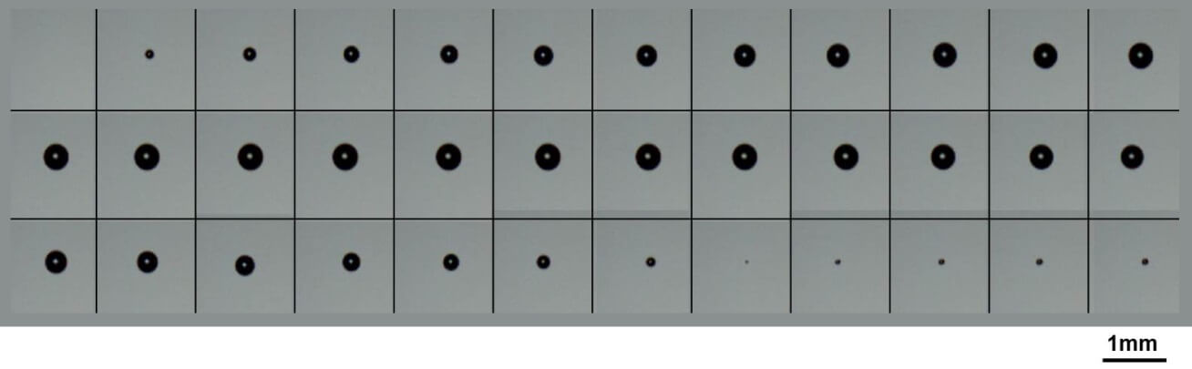
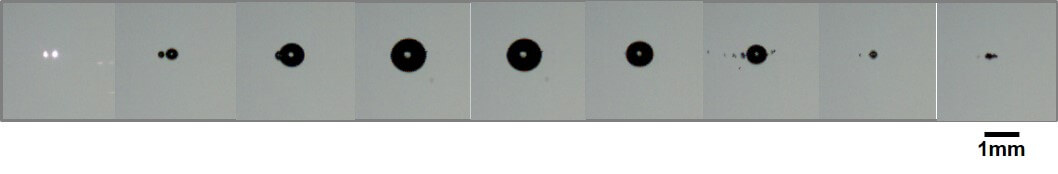
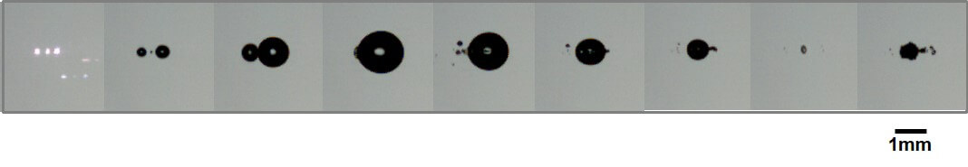
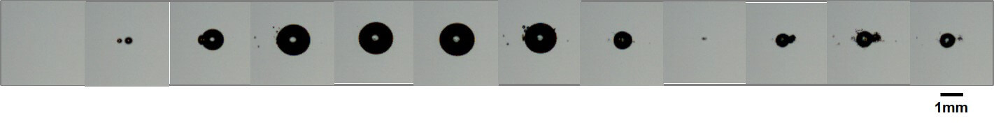
Fig. 5a Snapshots of bubble formation, growth and collapse for several cases of an L-bubble (a) and G-bubbles (b and c) with a laser energy input more than 7.5 mJ.
Frame rate is 86505, 84531and 53763 per second for (a), (b) and (c), respectively.

Reference
Jaekyoon Oh, Yungpil Yoo, Samsun Seung, Ho-Young Kwak. Laser-induced bubble formation on a micro gold particle levitated in water under ultrasonic field. Experimental Thermal and Fluid Science, volume 93 (2018) page 285–291.
Go To Experimental Thermal and Fluid Science