Significance
The integration of metallic implants into modern medicine—particularly in orthopedics and dentistry—has transformed the landscape of reconstructive and rehabilitative care. Devices composed of cobalt–chromium–molybdenum (CoCrMo) alloys have become central to joint replacement surgeries due to their excellent strength, wear resistance, and biocompatibility. Yet, despite these favorable characteristics, long-term success is far from guaranteed. Corrosion-induced degradation remains a quietly persistent issue that can compromise implant function, trigger adverse biological responses, and lead to premature failure. For clinicians and biomedical engineers alike, understanding the nuanced pathways through which corrosion evolves in vivo is no longer an academic exercise—it is a matter of patient safety and surgical outcome. A central challenge lies in the complex interplay between material microstructure and the physiological environment. Although CoCrMo alloys are known for forming protective oxide layers, localized corrosion still occurs, particularly at microscopic inhomogeneities such as grain boundaries or phase interfaces. These localized attacks are often exacerbated under pathological conditions, such as infection or inflammation, where the surrounding chemistry shifts in ways that accelerate electrochemical degradation. These environments cannot be approximated by standard in vitro tests alone; they demand a more tailored and physiologically relevant framework. One major gap in the existing literature is the limited understanding of how specific microstructural configurations—especially homogeneous versus banded arrangements—affect corrosion behavior under clinically relevant conditions. Previous investigations have highlighted the role of wear and fretting corrosion, but the direct influence of microstructure on electrochemical stability across different inflammatory states has not been systematically explored. It is this gap that motivated the present study. New research paper published in Surface and Coatings Technology and conducted by graduate student Maansi Thapa, Yani Sun, Bill Keaty, Professor Christos Takoudis, and Professor Mathew Mathew from the University of Illinois Chicago, researchers addressed this deficiency by isolating the effect of microstructure on corrosion susceptibility under three simulated biological scenarios: normal joint conditions, bacterial infection, and inflammatory stress. Their goal was compare two types of CoCrMo samples in a manner that reflects the biochemical realities faced by real implants inside the human body. By evaluating both electrochemical signatures and surface degradation patterns, the team aimed to uncover how subtle variations in internal structure can result in dramatically different outcomes once an implant is exposed to hostile biological environments.
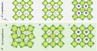
The researchers started by preparing two types of samples—one with a homogeneous microstructure and the other with a banded configuration. Each disk, precisely cut and polished to achieve a mirror-like finish, was then immersed in three distinct solutions meant to simulate environments an implant might encounter: a baseline representing normal physiological fluid, a second spiked with bacterial endotoxin to mimic infection, and a third containing hydrogen peroxide to replicate inflammatory stress. This trio of conditions was essential for capturing the range of biochemical challenges that orthopedic implants routinely face once inside the human body. With these test environments established, the researchers conducted electrochemical evaluations using a three-electrode system, relying on a saturated calomel reference electrode and a graphite counter. These tests, including cyclic polarization and electrochemical impedance spectroscopy (EIS), yielded detailed data about how readily each sample corroded. The findings were striking: across all three conditions, the homogeneous samples consistently showed lower corrosion current densities and more positive corrosion potentials. These metrics point to a surface that resists degradation more effectively, suggesting that a uniform grain structure provides fewer weak points for corrosive attack to take hold. The authors findings on EIS results further supported this. When the impedance data were modeled to extract polarization resistance and double-layer capacitance, the homogeneous microstructure again emerged as more resilient. Its surfaces displayed higher resistance to electrochemical change and lower capacitance, signaling a more stable oxide layer. Such a layer acts as a barrier, staving off the inward march of aggressive ions and prolonging the material’s integrity—an outcome that matters deeply in the context of implants expected to last decades. Moreover, surface-level observations offered another layer of evidence. Scanning electron microscopy revealed that the banded samples developed deeper pits and more pronounced grain boundary degradation, especially under infectious conditions. Meanwhile, profilometry showed that although surface roughness varied depending on the environment, the banded structure always bore more severe signs of localized damage. These visual insights confirmed what the electrochemical data hinted at: the banded microstructure, with its inherent variability, invited corrosion in ways that the homogeneous configuration simply did not.
In conclusion, the study of University of Illinois Chicago scientists successfully identified the difference in corrosion resistance between two microstructures and also exposes a quiet but critical factor in the long-term survival of biomedical implants by showing that homogeneous CoCrMo alloys withstand corrosion significantly better than their banded counterparts under a range of clinically relevant conditions, the authors indeed have illuminated a hidden determinant of implant longevity. This isn’t just a materials science insight—it has direct implications for patient outcomes, surgical planning, and the manufacturing standards that govern medical-grade alloys. We believe one of the most important takeaways lies in how subtle differences in internal structure can profoundly influence an alloy’s surface behavior once it enters the complex biochemical arena of the human body. What might appear under the microscope as a minor textural variation turns out to be a vulnerability. The banded microstructure, with its compositional and grain-boundary irregularities, becomes a foothold for corrosive reactions, especially when infection or inflammation changes the local environment. This is not merely a laboratory phenomenon—it mirrors real-world failures seen in revision surgeries, where corrosion products and ion release are known to trigger painful inflammatory responses and mechanical loosening. Additionally, by replicating these environments in vitro with thoughtful precision—introducing bacterial endotoxin to mimic infection and hydrogen peroxide to simulate inflammatory oxidative stress—the study closes the gap between bench research and clinical experience. It invites implant designers to re-evaluate assumptions about material uniformity and to recognize that corrosion resistance is not solely a function of bulk chemistry but is intimately tied to microstructural detail. The study as well for clinicians is equally meaningful. While device failure is often attributed to surgical technique or patient biology, this study suggests that the hidden architecture of the implant itself may be just as culpable. With clearer evidence linking microstructural uniformity to improved corrosion resistance, orthopedic and dental implant manufacturers may now feel pressure to demand tighter control over grain morphology during alloy processing. Standards might evolve. Surface treatments may be tailored more deliberately to accommodate microstructural characteristics rather than assumed homogeneity.


Reference
Maansi Thapa, Yani Sun, Bill Keaty, Christos Takoudis, Mathew Mathew, Corrosion risk analysis of CoCrMo alloy as a function of microstructure: Biomedical applications, Surface and Coatings Technology, Volume 497, 2025, 131757,