Significance Statement
Cell therapy is now being considered as the most direct method in restoring physiological functions of damaged organs and tissues. First generation cell therapy approach has been applied for several decades now despite the high risk of immune reactions. This has, therefore, indicated great potential for cell therapy. Researchers have developed 3-dimensional carriers as second generation cell therapy approach in a bid to protect the transplanted cells from immune reactions and enhance utilization efficiency. Among the developed 3-dimensional carriers, injectable hydrogels display intrinsic superiority to deliver and concentrate cells to the targeted areas while avoiding the risk of re-surgery.
However, there’s still a huge gap between successful applications of the hydrogels in cell therapy. The major limitations are that physical hydrogels depend on changes in physical conditions which might affect cell viability. Gelation of these hydrogels after injection are hard to control. Chemical hydrogels generated via covalent bonding have complex chemical reactions which have high toxicity, therefore limiting in vivo applications. In artificial polymer hydrogels, growth factors are needed to initiate proliferation of the cells, which could cause some risks to the surrounding cells.
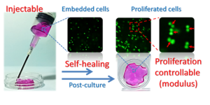
Developing a gelation controllable, biodegradable and biocompatible hydrogel that can prompt high viability and proliferation of the encapsulated cells without the use of growth factors after injection is of ultimate importance. Yongsan Li and colleagues developed, for the first time, an injectable, self-healing dynamic hydrogel implementing a biocompatible glycol chitosan and poly (ethylene glycol) as gelators. Their work is published in peer-reviewed journal, Colloids and Surfaces B.
The authors prepared a series of hydrogels by dissolving glycol-chitosan in deionized water and then mixing it with varying concentrations of dibenzaldehyde-terminated poly(ethyleneglycol) solution. They came up with three hydrogels with different moduli. The team prepared a 400 uL of hydrogel and after complete gelation, the hydrogel was cut into approximately 25 pieces to imitate the damage of hydrogels immediately after injection. Afterwards, the researchers monitored the self-healing behavior of the gels.
The authors recorded storage moduli of 0.9kPa (soft), 2.0kPa (medium) and 4.7kPa (stiff) for hydrogels synthesized with 1.5% glycol chitosan and, respectively, 1%, 2%, and 4% of (cross-linker) dibenzaldehyde-terminated poly(ethyleneglycol). The modulus of the stiffest hydrogel was found to be near that of subcutaneous tissue.
The authors observed that the chitosan-based dynamic hydrogel would heal after injection. They implemented a rheology test in a bid to trace modulus change according to time increase. The storage modulus reduced after hydrogel damage and them increased with time until the value was the same as that of a pristine hydrogel. This was a good indicator of a self-healing process. The researchers observed that the soft hydrogel self-healed to the original modulus in 40 minutes while for the stiff hydrogel took 84 minutes. Control and middle groups healed in relatively the same time indicating that the addition of poly(ethyleneglycol) had no impact on the hydrogel modulus.
The authors prepared chitosan-based hydrogels with varying storage moduli. They came up with correlation between the hydrogel modulus and the proliferation rate of the embedded cells, which was a facile method to tame cell proliferation in the hydrogel without using a growth factor. The cells possessed high viability and maintained high proliferation rate in the hydrogels indicating that the self-healing hydrogel would deliver the implanted cells to the targeted spot and enhance curing by allowing for in-situ proliferation of the encapsulated cells.
Chitosan-based dynamic hydrogel appeared to be a promising candidate for incubating functional cells for cell therapy. However, a balance between the negative shearing force and positive stiffness factor must be taken into account. The proposed chitosan-based hydrogel will definitely enhance biological usage for chemically cross-linked hydrogels in areas of cell delivery.

Reference
Yongsan Li1,2, Yingwei Zhang1, Feng Shi1, Lei Tao2, Yen Weib2, and Xing Wang1. Modulus-regulated 3D-cell proliferation in an injectable self-healing hydrogel. Colloids and Surfaces B: Biointerfaces, volume 149 (2017), pages 168–173.
[expand title=”Show Affiliations”]- Beijing Laboratory of Biomedical Materials, Beijing University of Chemical Technology, Beijing 100029, PR China
- The Key Laboratory of Bioorganic Phosphorus Chemistry & Chemical Biology (Ministry of Education), Department of Chemistry, Tsinghua University, Beijing 100084, PR China
Go To Colloids and Surfaces B: Biointerfaces