International Journal of Adhesion and Adhesives, Volume 42, April 2013, Pages 21-29.
H.D. Ford, R.P. Tatam
Department of Engineering Photonics, School of Engineering, Cranfield University, Cranfield, Bedford MK43 0AL, United Kingdom
Abstract
Cross-correlation of optical coherence tomography (OCT) images has been applied to internal imaging of PVA and two-part epoxy adhesives during cure, providing information on relative viscosity at different positions within the sample volume. Spatial resolution of a few micrometres is obtained in the original OCT images, and a few tens of micrometres in the correlation images, enabling the progress of cure to be mapped in fine detail within a small cure volume. Evidence of phase separation is seen in the OCT and correlation images of a partially-cured PVA emulsion. Mixing structure and regions of poor cure can be observed in a poorly-mixed two-part epoxy.
Additional information
Correlation techniques, applied to images obtained using optical coherence tomography (OCT), provide a non-contact method of monitoring relative viscosity, and hence degree of cure, in semi-transparent adhesives. In contrast to many existing methods for cure-monitoring, the OCT technique described in this paper provides spatially-resolved information, with a spatial resolution of the order of 10s of micrometres, allowing the user to identify regions of slower or faster cure within a 2D image corresponding to a selected vertical slice through the sample.
Figure Legend
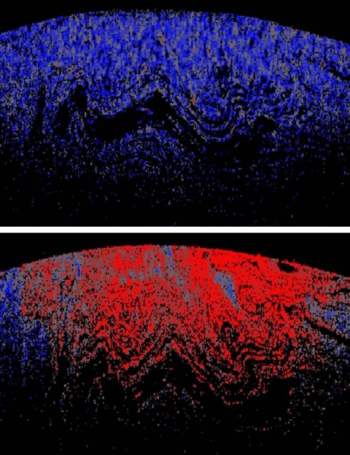
Images (representing a region 3mm across x 1.6 mm deep) show correlation OCT images for a sample of poorly-mixed two-part ‘Araldite Rapid’ epoxy resin. Top image: 2 minutes after mixing; 20 s correlation interval. Lower image: I hour after mixing; 2 minute correlation interval. Blue represents a low correlation coefficient (low viscosity) and red a high correlation coefficient (high viscosity), with grey colouration in the mid-range.
In the upper image, the viscosity is uniformly low. However, in the lower image, although viscosity has generally increased, as much of the sample has undergone a significant degree of cure, pockets of poorly-cured mixture can be identified within the image, suggesting where regions of weakness might be found in the final work piece.