J. Appl. Phys. 114, 034901 (2013);
Kin Mun Wong, S. M. Alay-e-Abbas, Yaoguo Fang, A. Shaukat,Yong Lei.
Institut fUr Physik & IMN MacroNano® (ZIK), Technische Universität Ilmenau, Prof. Schmidt-Str. 26, Ilmenau 98693, Germany and
Department of Physics, GC University Faisalabad, Allama Iqbal Road, Faisalabad 38000, Pakistan and
Department of Physics, University of Sargodha, 40100 Sargodha, Pakistan
Abstract
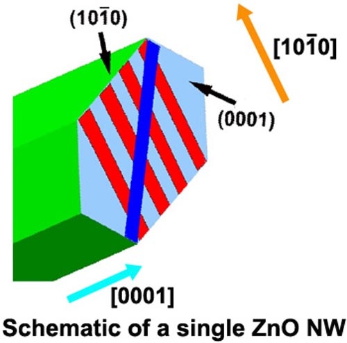
A qualitative approach using room-temperature confocal microscopy is employed to investigate the spatial distribution of shallow and deep oxygen vacancy (VO) concentrations on the polar (0001) and non-polar (10Ī0) surfaces of zinc oxide (ZnO) nanowires (NWs). Using the spectral intensity variation of the confocal photoluminescence of the green emission at different spatial locations on the surface, the VO concentrations of an individual ZnO NW can be obtained. The green emission at different spatial locations on the ZnO NW polar (0001) and non-polar (10Ī0) surfaces is found to have maximum intensity near the NW edges, decreasing to a minimum near the NW center. First-principles calculations using simple supercell-slab (SS) models are employed to approximate/model the defects on the ZnO NW (10Ī0) and (0001) surfaces. These calculations give increased insight into the physical mechanism behind the green emission spectral intensity and the characteristics of an individual ZnO NW. The highly accurate density functional theory (DFT)-based full-potential linearized augmented plane-wave plus local orbitals (FP-LAPW + lo) method is used to compute the defect formation energy (DFE) of the SSs. Previously, using these SS models, it was demonstrated through the FP-LAPW + lo method that in the presence of oxygen vacancies at the (0001) surface, the phase transformation of the SSs in the graphite-like structure to the wurtzite lattice structure will occur even if the thickness of the graphite-like SSs are equal to or less than 4 atomic graphite-like layers [Wong et al., J. Appl. Phys. 113, 014304 (2013)]. The spatial profile of the neutral VO DFEs from the DFT calculations along the ZnO [0001] and [10Ī0] directions is found to reasonably explain the spatial profile of the measured confocal luminescence intensity on these surfaces, leading to the conclusion that the green emission spectra of the NWs likely originate from neutral oxygen vacancies. Another significant result is that the variation in the calculated DFE along the ZnO [0001] and [10Ī0] directions shows different behaviors owing to the non-polar and polar nature of these SSs. These results are important for tuning and understanding the variations in the optical response of ZnO NW-based devices in different geometric configurations.
© 2013 Author(s).All article content, except where otherwise noted, is licensed under a Creative Commons Attribution 3.0 Unported License.
Additional Information
It is well known that the ZnO nanowire (NW) contain a large density of intrinsic defects such as oxygen deficiencies or vacancies (VO), which influence their electronic and optical properties as well as their performance in device applications. Hence understanding the spatial distribution of the shallow intrinsic surface defects on the nanostructure’s surfaces and deep intrinsic bulk defects plays a crucial role in understanding and tailoring the electronic properties of the NW based device. The ZnO (0001) and (10Ī0) surfaces are the most important among the different surface structures of ZnO as they occur predominantly due to their relative stability. In the article, from multiple arguments, it is likely that the green luminescence emitted by the synthesized NW arrays during the confocal photoluminescence (CPL) microscopy measurements originates from neutral oxygen vacancies. In addition, the luminescence intensity or the magnitude of the measured CPL spectra is also directly correlated with the concentration of VO in the ZnO NWs. Therefore the CPL measurements can provide a direct and non-destructive qualitative measurement of the directional dependence of the VO concentration in the ZnO NWs. The spatial profile of the luminescence intensity indicates that the VO concentration is largest near the edges of the (0001) and (10Ī0) surfaces of the NWs and the VO concentrations decreases significantly in the interior parts of the NWs
In order to gain insight into the physical mechanisms that govern the confocal optical properties of the ZnO NWs in the [0001] and [10Ī0] directions, density functional theory calculations using the full-potential linearized augmented plane-wave plus local orbitals method was also utilized. It was demonstrated that the supercell-slab (SS) models in the surface simulations was suitable as an approximation of the ZnO NW cross sections where the confocal measurements are taken. The calculations show that the spatial electronic profile of the neutral VO formation energy in the [0001] and [10Ī0] directions is lower near the boundary and increases to bulk-like values in the SS center, which explains the behaviour of the spatial profile of the CPL measurements. Using this combined experimental and theoretical approach, a better qualitative understanding of the spatial distribution of the surface and deep VO in the ZnO NWs was achieved, thus leading to a more efficient functionalization and improved integration of the ZnO NW in future nanodevices for better performance.