The Journal of Supercritical Fluids, Volume 82, 2013, Pages 50-55.
Zhe Xing, Mouhua Wang, Guohao Du, Tiqiao Xiao, Weihua Liu, Dou Qiang, Guozhong Wu.
Shanghai Institute of Applied Physics, Chinese Academy of Sciences, Shanghai 201800, China and
University of Chinese Academy of Sciences, Beijing 100049, China.
Abstract
Non-destructive X-ray microtomography at a third generation synchrotron facility was applied to analyze the cell morphology of microcellular polystyrene (PS)/polyethylene (PE) alloy foams. This method, differing from the observation of cross section of cell by SEM, enables one to observe a complete cell structure in the polymer foam. PS/PE foams were prepared using a supercritical CO2 foaming process. A styrene–ethylene–butylene–styrene (SEBS) copolymer was used as the compatibilizer of PS and PE to improve the cell morphology. The effects of PS/PE composition and foaming conditions (temperature and pressure) on the cell structure of foams were investigated in detail. The optimal SEBS content for the foaming of PS/PE (70:30) alloys was found to be 5 wt%. The cell size and cell density were also dependent on the foaming temperature and the saturation pressure.
Additional Information
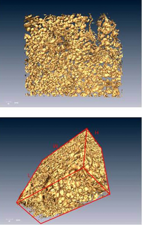
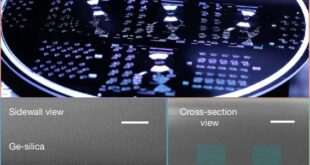
We have prepared PS/PE alloy foams using a scCO2 foaming process and analyzed the foaming specimens using X-ray microtomography (XMT) at the X-ray imaging and biomedical applications beamline (BL13W1) of Shanghai Synchrotron Radiation Facility (SSRF), which is now the largest scientific facility in China. The BL13W1 dedicates to development and applications of cutting-edge X-ray imaging techniques in different scientific research fields. Since the X-ray flux of synchrotron radiation (SR) is several orders of magnitude higher than that of conventional X-ray tube, SR imaging has much higher spatial and contrast resolutions. The SR imaging can be utilized to obtain in-situ, non-destructive, high-resolution and three-dimensional images of various materials, including low-Z materials like soft tissue and polymer. As an in-house work, it is our motivation to characterize the micro-structure of polymer foams at the beamline of SRRF, and make a clear comparison with the observation with scanning electron microscopy (SEM).
Compared to the observation of cross-section cell structure of polymer foam by SEM, XMT at SSRF can be utilized to reconstruct high-resolution interior structural details of polymer foam, through comparison of a three dimensional intensity distribution of a specimen’s X-ray absorption. XMT overcomes drawbacks of current imaging techniques, such as light microscopy and SEM. For example, destruction of cells due to the cutting of foam to expose the cross-section to observation can alter the structural features in SEM. SR XMT is non-destructive and can provide accurate three-dimensional structure information of polymer foams, the important microstructural features like cell size distribution, degree of interconnectedness between cells, average cell wall thickness and open cell volume fraction can be directly measured with high accuracy. Therefore, SR XMT is an important tool that holds great potential for estimation of interior properties of polymer foam materials. Below are snapshots of reconstructed 3D images of the PS/PE alloy foam prepared by scCO2 foaming process, for a better understanding of the three dimensional features of foam.