Applied Surface Science, Volume 275, 15 June 2013, Pages 409-412.
K. Oshida, M. Murata, K. Fujiwara, T. Itaya, T. Yanagisawa, K. Kimura, T. Nakazawa, Y.A. Kim, M. Endo, B.-H. Kim, K.S. Yang.
Nagano National College of Technology, Nagano 381-8550, Japan &
GSI Creos Corporation, Kanagawa 210-0855, Japan &
Shinshu University, Nagano 380-8553, Japan &
Chonnam National University, Gwangju 500-757, Republic of Korea
Abstract
Transmission electron microscopy (TEM) is one of the highest resolution analysis methods of materials. The three dimensional recognition of the materials is difficult by TEM because the observation data is projection images through the materials. In this study, space structure of carbon nanotubes loaded with metal particles was analyzed by three dimensional TEM (3D-TEM) 0005 and 0010. The nano structured carbons are also observed by high resolution transmission electron microscopy (HRTEM) with Cs corrector. Cup-stack type carbon nanotubes (CSCNTs) loaded with Pt particles (2–3 nm in diameter) prepared by GSI Creos Corporation were analyzed by these methods. Pt particles are bound selectively to the edges of hexagonal carbon layers of inside and outer surface of CSCNTs efficiently and can be expected to work well as catalysts of electrodes of fuel cell. It is sometimes difficult that the nano sized area is analyzed by selected area electron diffraction (SAD) because the selected area aperture cannot be so small. The HRTEM and image processing technique give similar results of SAD when it works and revealed to be useful to analyze nano structured carbons.
Additional Information
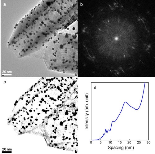
Cupstack type carbon nanotubes (CSCNTs) loaded with Pt particles were observed by high resolution transmission electron microscopy (HRTEM) with Cs corrector and three dimensional TEM (3D-TEM). The IRTEM images were analyzed by image processing. The HRTEM image is shown in figure (a). Figure (b) shows power spectrum image of (a) obtained by 2 dimensional Fourier transform (2D-FFT). The graph as shown in figure (d) was obtained by integration of the power spectrum around the center point of it. The distribution of distances among the Pt particles is estimated to less than 25nm by analysis of figure (d). The 2D-FFT data corresponding to less than 25nm was extracted and the real space image was reconstructed by 2 dimensional inverse fast Fourier transform (2D-IFFT). The binary image of the real space image is shown in figure (c). The Pt particles are well extracted by this series of operation. The result reveals that the peaks in figure (d) indicate the distribution of Pt particles. Pt particles are bound selectively to the edges of hexagonal carbon layers of inside and outer surface of CSCNTs efficiently and can be expected to work well as catalysts of electrodes of fuel cell. The analysis method using 3D-TEM together HRTEM and image processing is useful to understand the nano-structure of materials accurately.