European Polymer Journal, 18 March 2013.
Aysegul Cumurcu, Joost Duvigneau, Ian D. Lindsay, Peter Schon, G. Julius Vancso
Materials Science and Technology of Polymers, MESA+ Institute for Nanotechnology, University of Twente, Enschede NL-7500, The Netherlands.
Nanophysics and Soft Matter Group, H.H. Wills Physics Laboratory, University of Bristol, Tyndall Avenue, Bristol BS8 1TL, United Kingdom.
Abstract
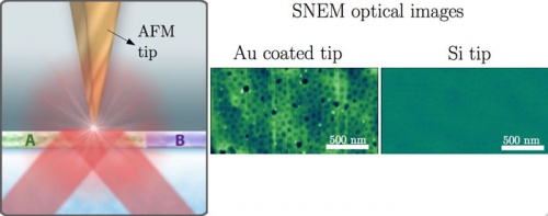
Scanning near field ellipsometric microscopy (SNEM) was used to simultaneously obtain optical images and tapping mode topography images of the microphase separated morphology of PS-b-P2VP block copolymer thin films. Optical images revealed a spatial resolution well below the diffraction limit. The SNEM setup used consisted of an AFM and an ellipsometer that were commercially available and that did not require major changes in their design to build the hybrid microscope. The observed increase in optical contrast for gold coated tips compared to silicon AFM probe tips was in qualitative agreement with the calculated increase in scattering amplitude according to the point dipole model for a gold AFM probe tip compared to a bare silicon AFM probe tip of the same size. The dielectric constant difference between the two blocks of the diblock copolymer was increased by selectively staining the P2VP block with iodine vapor. This resulted in an increase in the optical contrast between the PS and P2VP domains. Furthermore, the decrease in optical contrast as a function of increasing tip-sample separation was studied. It was observed that at 50 nm tip sample separation the optical contrast was significantly reduced. The non-linear decay of the near-field amplitude signal as a function of the tip-sample separation calculated with the point dipole model supported this experimental result. The use of tapping mode in SNEM opens novel opportunities to study soft matter down to the macromolecular level.
Additional Information
In the past 20 years, the atomic force microscope (AFM) has evolved into a powerful tool for the nanoscale investigation of polymeric materials down to the single macromolecule level. Although AFM can provide images with nanometer spatial resolution on topography and mechanical properties of heterogeneous materials, there is a fundamental need to obtain further material specific information, for instance optical properties with nanoscale resolution. In this regard the integration of an AFM with optical instrumentation into hybrid devices has opened novel avenues for nanoscale material characterization.
We recently introduced a hybrid setup called scanning near field ellipsometric microscope (SNEM) composed of an AFM and an ellipsometer. This approach relies on recording changes in the state of polarization of the electromagnetic field by a vibrating AFM probe in the proximity of the specimen surface. For excitation of gold-coated probes, field enhancement at the tip apex would be expected to occur both as a result of the increasing confinement of the surface charge density at the sharp tip apex, the so-called “lightning-rod effect” and due to the resonant excitation of localized surface plasmon modes of the metalized probe tip. SNEM was used to simultaneously obtain optical images and topography images of the microphase separated morphology of block copolymer films. Optical images of the block copolymer films revealed a spatial resolution well below the diffraction limit. Here, we report on the investigation of the effect of the tip coating, tip-sample separation distance and the dielectric constant difference in the sample on the optical contrast. These results have improved the understanding of the optical contrast mechanism of SNEM. Calculations of the point dipole model were in qualitative agreement with the experimental results.