 Advances in Engineering Advances in Engineering features breaking research judged by Advances in Engineering advisory team to be of key importance in the Engineering field. Papers are selected from over 10,000 published each week from most peer reviewed journals.

International Journal of Adhesion and Adhesives, Volume 42, April 2013, Pages 21-29.
H.D. Ford, R.P. Tatam
Department of Engineering Photonics, School of Engineering, Cranfield University, Cranfield, Bedford MK43 0AL, United Kingdom
Abstract
Cross-correlation of optical coherence tomography (OCT) images has been applied to internal imaging of PVA and two-part epoxy adhesives during cure, providing information on relative viscosity at different positions within the sample volume. Spatial resolution of a few micrometres is obtained in the original OCT images, and a few tens of micrometres in the correlation images, enabling the progress of cure to be mapped in fine detail within a small cure volume. Evidence of phase separation is seen in the OCT and correlation images of a partially-cured PVA emulsion. Mixing structure and regions of poor cure can be observed in a poorly-mixed two-part epoxy.
Additional information
Correlation techniques, applied to images obtained using optical coherence tomography (OCT), provide a non-contact method of monitoring relative viscosity, and hence degree of cure, in semi-transparent adhesives. In contrast to many existing methods for cure-monitoring, the OCT technique described in this paper provides spatially-resolved information, with a spatial resolution of the order of 10s of micrometres, allowing the user to identify regions of slower or faster cure within a 2D image corresponding to a selected vertical slice through the sample.
Figure Legend
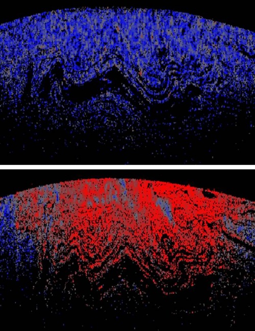
Images (representing a region 3mm across x 1.6 mm deep) show correlation OCT images for a sample of poorly-mixed two-part ‘Araldite Rapid’ epoxy resin. Top image: 2 minutes after mixing; 20 s correlation interval. Lower image: I hour after mixing; 2 minute correlation interval. Blue represents a low correlation coefficient (low viscosity) and red a high correlation coefficient (high viscosity), with grey colouration in the mid-range.
In the upper image, the viscosity is uniformly low. However, in the lower image, although viscosity has generally increased, as much of the sample has undergone a significant degree of cure, pockets of poorly-cured mixture can be identified within the image, suggesting where regions of weakness might be found in the final work piece.